
Image-guided systems function like GPS (Global Positioning System) systems in a patient’s head. They help confirm the location of critical structures when the interior of the nose and sinuses is distorted by unusual anatomy or prior surgery.
How Image-Guided Surgery Works
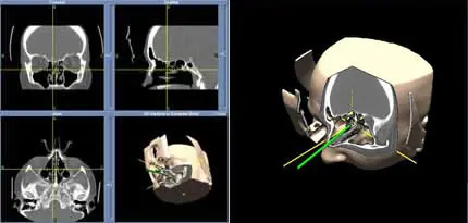
A CT scan of the sinuses is performed using a specific navigation system protocol. For some systems, a special mask or markers are placed on your face during the scan to serve as reference points. The CT scan is transferred to a disk, which is then loaded into the image-guidance computer.
During surgery, a detection array or a mask is placed on the patient’s head. The CT scan images loaded into the system are then calibrated to the patient’s anatomy using preset reference points, such as the mask, markers or specific anatomic points on the face. The computer can then track the position of the sinus surgery instruments by integrating information from the patient’s pre-set reference points and comparing it with the CT scan map.
When Image-Guided Surgery Is Needed
Image-guided surgery is not necessary during all ENT procedures. It is typically not needed for patients undergoing their first sinus surgery or those with straightforward anatomy.
Image guidance is most helpful for patients who have:
- Have previously undergone sinus surgery
- Have complicated anatomy in the sinuses
- Cases of benign tumor growths, such as inverted papilloma
- Cerebrospinal fluid leaks
Our experts will determine if image-guided surgery is a suitable option for you during your procedure. We are here to answer any questions you have tos ensure you feel confident about the process.
Call California Sinus Centers for more information or to schedule an appointment.